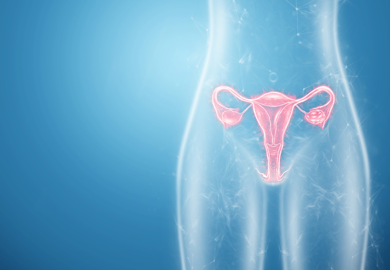

A vaginal septum is an abnormality that causes the vagina to be divided into two canals. The septum is formed during fetal development and can cause problems in the future.
Causes of a Vaginal Septum
A vaginal septum forms when a fetus is developing in the mother’s womb. The risk factors for this condition are unknown.
Female reproductive organs begin to develop at about six weeks after fertilization. In normal development, Müllerian ducts fuse together centrally, with some tissue being used to form the uterus and fallopian tubes. Other tissues are used to form the vaginal walls of a single vaginal canal.
Any mutation that occurs at this time is called a Müllerian anomaly. This can lead to a vaginal septum. Women with a vaginal septum may have other internal malformations, such as:
- Skeletal anomalies (bone)
- Renal anomalies (kidney)
- Anorectal anomalies (anus or rectum)

Types of Vaginal Septum
The three types of vaginal septum are classified based on shape, thickness, and location.
- Longitudinal vaginal septum: This septum runs vertically through the vagina, dividing the vagina into two separate canals. Most women with longitudinal vaginal septum don’t have symptoms, but a physician may identify it during a routine pelvic exam.
- Transverse vaginal septum: This septum runs horizontally across the vagina. The transverse vaginal septum can be complete, causing full blockage of the upper vagina, or partial, only partially blocking the vagina. A complete blockage of the vagina can cause missed menstrual cycles, swelling of the upper abdomen, and pain during sexual intercourse.
- Obstructed hemi vagina: An obstructed hemi vagina has a septum that blocks and divides half of the vagina. Women typically have a normal menstrual cycle in one half of the vagina, and blockages on the other side. Many women may also have a didelphic — or double uterus — and experience extreme menstrual pain.
Diagnosis of a Vaginal Septum
To diagnose a vaginal septum, the physician will review the patient’s symptoms and medical history, perform a pelvic exam, and ask the patient to get an MRI done.
Women with a septum that doesn’t block the vagina or cause any symptoms may not need treatment. But women with ab obstructive septum may experience issues with tampon use, sexual intercourse, and natural childbirth.
Types of Surgical Resection for a Vaginal Septum
The type of surgical resection done to treat a patient will depend on the kind of vaginal septum they have.
Resection of a longitudinal vaginal septum: In patients with a vagina divided into two separate canals, the physician will remove the septum and the abnormal tissue surrounding it, leaving the vagina as a single canal.
Resection of a transverse vaginal septum: This type of resection is more complex, requiring other interventions to be made in advance. For example, if the septum blocks the flow of blood during menstruation, the physician may first drain the obstructed blood and then remove the transverse septal tissue.
Resection of an obstructed hemi vagina: The physician will remove vaginal septum tissue to create a single canal and remove menstrual blood blockages.
