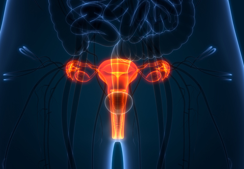

Congenital müllerian anomalies occur when müllerian ducts don’t develop properly, disrupting development of the entire reproductive system, including the uterus, cervix, fallopian tubes, and upper two-thirds of the vagina.
Symptoms of Congenital Müllerian Anomalies
Women with abnormalities in the müllerian ducts may experience the following symptoms:
- Infertility
- Recurrent miscarriage
- Pelvic pain
- Preterm labor
- Lack of monthly periods (amenorrhea)
- Difficulty with tampon use or intercourse
Delays in accurate diagnosis and treatment can lead to chronic pelvic pain, increased retrograde menstruation, endometriosis, infertility, and missed school/work days.

Types of Müllerian Anomalies
There are about 10 different types of müllerian anomalies affecting the uterus and cervix:
1. Müllerian agenesis. When a female is born without a uterus, typically due to a broader condition involving several abnormalities of the reproductive system.
2. Unicornuate uterus. When a female only has half a uterus, shaped like a banana.
3. Didelphys uterus. When a female has a double uterus, shaped like a banana, due to improper formation during fetal development.
4. Bicornuate uterus. A heart-shaped uterus with an indentation at the top and one cervical opening.
5. Septate uterus. A heart-shaped inner cavity of the uterus.
6. Cervical agenesis. Missing cervix.
7. Cervical duplication. Two cervixes.
8. Cervical hypoplasia. An abnormally small cervix.
9. Vaginal septum. Longitudinal dividing the vagina in half, or a transverse septum that can block menstrual outflow.
10. Obstructed uterine horn. When one piece of a uterus forms without the cervix or vagina attached.
The most common müllerian anomaly is vaginal septum. In this case, there can be a lateral or vertical fusion issue. A lateral fusion can lead to a unicornuate uterus.
Symptoms will depend on where the septum is located and whether the uterus has an endometrial lining. If the uterine remnant is disconnected from the vagina and has an endometrial lining, then menstrual outflow will be blocked, causing pain. Women may also develop pelvic masses if the uterine remnant or fallopian tube fill with blood.
How Congenital Müllerian Anomalies Are Diagnosed
During a medical appointment, the gynecologist will note patient symptoms and perform a pelvic exam looking at the external and internal female organs. Imaging tests will then be performed to further analyze the patient’s condition. Tests may include:
- Pelvic ultrasound
- Pelvic magnetic resonance imaging (MRI)
- Hysteroscopy, which involves inserting a tiny telescope through the vagina into the uterus to view and treat abnormal areas
- Hysterosalpingography, or an X-ray involving a dye
- Vaginoscopy, which involves inserting a tiny telescope into the vagina to view abnormal areas
- Laparoscopy, which involves inserting a thin tube into the abdomen with a high-intensity light and high-resolution camera to view abnormal areas
Treatment For Congenital Müllerian Anomalies
Treatment for these anomalies typically involve reducing the risk of pregnancy complications, including miscarriages, early deliveries, and breech births. Women without a uterus have limited treatment options, but may undergo uterine transplants. The right treatment and surgical approach will depend on the unique type of abnormality.
Women with pelvic pain or irregular periods caused by müllerian anomalies may undergo surgery that can range from vaginal surgery (such as septum removal) to a minimally invasive laparoscopic procedure. In rare cases, a hysterectomy may be the only option.
